Research
Some regions of the human genome are simply hard to copy.
Dense in repetitive sequence, prone to folding into unusual structures and often carrying long, actively transcribed genes, these fragile sites stall the replication machinery and break under stress that the rest of the genome absorbs without incident. Our lab studies why.
The projects below start from a shared observation: when damage lands in the same place again and again, the explanation is usually that the place itself is difficult, not that the damage was aimed there.
Different diseases, one underlying question — what makes a stretch of DNA vulnerable, and why in these cells and not others.
Six steps joined left to right by arrows: genotoxic insult (dietary dyes, WTC dust, protein deficiency, viral infections); DNA damage and stalled forks; damage tolerance versus DNA repair, the fork in the road; genomic scars (breaks, micronuclei, signatures); pre-cancer (clonal expansion); and cancer. Projects 1, 2 and 3 sit under the first step, project 4 under the second and project 5 under the third; project 2 again under pre-cancer; and a branch from pre-cancer leads to early detection, the therapeutic window.
Genotoxic insult
dietary dyes, WTC dust, protein deficiency, viral infections
DNA damage & stalled forks
Damage tolerance vs. DNA repair
the fork in the road
Genomic scars
breaks, micronuclei, signatures
Pre-cancer
clonal expansion
Cancer
Dietary Azo Dye Toxicity: Mutagenesis & Carcinogenesis
Red 40 and Yellow 6 are the two most heavily used azo dyes in the U.S. food supply. Gut bacteria cleave them into reactive aromatic amines that attack DNA.
The Fanconi anemia pathway is the BRCA pathway: an early-warning readout for the 1 in 300–400 people carrying a BRCA variant.
- Supported by
- New Jersey Center for Pediatric Cancer & Blood Disorders Research.
Four steps joined by arrows: Red 40 and Yellow 6, the two most heavily used azo dyes in the U.S. food supply, in cereals, candy, sports drinks, snacks and medicines; gut bacteria cleave them into reactive aromatic amines that attack DNA; DNA damage, with a dysregulated FA/BRCA DNA repair pathway; and the open question, do those lesions push blood stem cells toward leukemia? From the question two arrows fan out to the disease sites, drawn as end states: blood and bone marrow, leukemia and marrow failure; and breast and ovary, through the shared BRCA genes, with the whole body susceptible.
Red 40 and Yellow 6
the two most heavily used azo dyes in the U.S. food supply — in cereals, candy, sports drinks, snacks, even medicines
Gut bacteria cleave them
into reactive aromatic amines that attack DNA
DNA damage
dysregulated FA/BRCA DNA repair pathway
Do those lesions push blood stem cells toward leukemia?
the question we are asking
Blood and bone marrow
leukemia and marrow failure
Breast and ovary
through the shared BRCA genes — and the whole body is susceptible
The same genes, two clinical names
- BRCA2
- = FANCD1
- BRCA1
- = FANCS
- PALB2
- = FANCN
- BRIP1
- = FANCJ
- RAD51C
- = FANCO
Breast and ovarian cancer genes; two damaged copies of each cause a Fanconi anemia subtype.
Who this reaches
- 1 in 300–400
- carry a BRCA1/2 variant
- 1 in 40
- of Ashkenazi Jewish ancestry
- 9.2 million
- New Jerseyans we serve
Our catchment area is the entire state, including all 21 counties. Breast cancer is a named Rutgers Cancer Institute priority, and New Jersey incidence sits significantly above the U.S. average.
Why this is an equity-friendly intervention
- No cost
- reading a label is free
- No gatekeeper
- no insurance, referral or specialist visit
- No clinic
- a family can act on it tomorrow
Dye-heavy processed foods are cheapest and most available in New Jersey’s food-insecure neighborhoods, where cancer mortality is already highest.
9/11 World Trade Center Exposure: From Dust to Leukemia
First responders carry elevated rates of premalignant and malignant blood cells decades after the collapse. Which mutational processes does a complex exposure leave behind, and why do they land at the same few loci every time?
Four steps joined by arrows: World Trade Center dust, the exposure first responders took at the collapse in 2001; somatic mutation at a few loci — DNMT3A, TET2, ASXL1, JAK2, PPM1D and SF3B1 — in blood stem cells, again and again; clonal hematopoiesis, elevated rates of premalignant and malignant blood cells decades later; and the open question, are these regions undiscovered fragile sites, given their stress-dependent hypermutability, cell-type specificity and an expressed gene. From the question two arrows fan out to the end states: leukemia, for which clonal hematopoiesis is a recognized precursor, and cardiovascular disease, because the clone’s differentiated cells circulate through the whole body.
World Trade Center dust
first responders at the collapse, 2001
Somatic mutation at a few loci
DNMT3A, TET2, ASXL1, JAK2, PPM1D, SF3B1 — in blood stem cells, again and again
Clonal hematopoiesis
elevated rates of premalignant and malignant blood cells, decades after the collapse
Are these regions undiscovered fragile sites?
stress-dependent hypermutability, cell-type specificity, an expressed gene — the three hallmarks
Leukemia
clonal hematopoiesis is a recognized precursor state
Cardiovascular disease
the clone’s differentiated cells circulate through the whole body
First responders to the World Trade Center attacks carry elevated rates of premalignant and malignant blood cells, decades after the collapse. That fact is established. What it has not yet told us is how — which mutational processes a complex environmental exposure actually leaves behind in the blood, and why they land where they do.
Rutgers hosts a World Trade Center Health Program Clinical Center of Excellence, so the population this research concerns is one our own institution monitors and treats.
Our retrospective study of this cohort points toward somatic hypermutation at genes associated with clonal hematopoiesis. In clonal hematopoiesis, somatic mutations at a defined set of loci give hematopoietic stem cells a fitness advantage and allow them to expand clonally. It is not always harmful, but it is a recognized precursor state for leukemia, and because differentiated blood cells circulate throughout the body, it also carries elevated cardiovascular risk.
These mutations recur at a strikingly small number of loci — DNMT3A, TET2, ASXL1, JAK2, PPM1D, SF3B1 — and are thought to accumulate in response to endogenous and exogenous stress across a lifetime. What remains unexplained is why these particular loci. Why does a diffuse, whole-body exposure converge on the same few regions, in the same cell type, again and again? The recurrence is usually treated as a given rather than as something requiring explanation.
Beyond clonal hematopoiesis itself, the answer may help explain why individuals with genome instability disorders such as Fanconi anemia are predisposed to blood cancers.
- Supported by
- W81XWH-21-1-0935 and U01OH012271 (co-investigator).
EP300 Loss in Adult T-cell Leukemia: Finding the Therapeutic Window
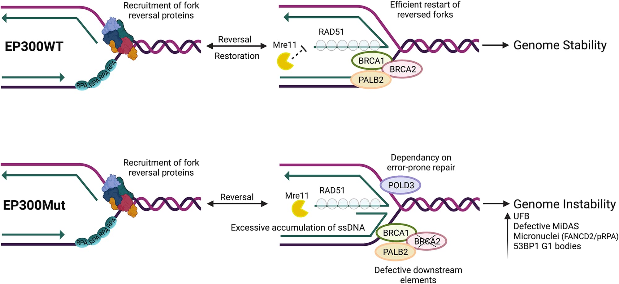
Targeting BRCA2 loss — EP300 loss leads to BRCA2 protein loss; cells behave like BRCA-deficient tumors, sensitive to PARP, REV1 and Pol θ inhibitors.
Mitochondrial dysfunction: how chronic replication stress reshapes mitochondrial function and cytosolic DNA sensing.
- Supported by
- R01CA307099 and R01CA266847 (co-investigator).
Three steps joined by arrows: EP300 loss in adult T-cell leukemia; BRCA2 protein loss, and with it defective replication fork protection; and cells that behave like BRCA-deficient tumors under chronic replication stress. An arrow leads from there to the therapeutic window the project’s title names, outlined: sensitivity to PARP, REV1 and Pol θ inhibitors.
EP300 loss
in adult T-cell leukemia
BRCA2 protein loss
and with it, defective replication fork protection
Cells behave like BRCA-deficient tumors
under chronic replication stress
Sensitive to PARP, REV1 and Pol θ inhibitors
the therapeutic window
Who this reaches
- 14.1 per million
- incidence among non-Hispanic Caribbean-born U.S. residents
- 0.4 per million
- among those born in the U.S. or Canada
- 23.8%
- five-year survival among Caribbean-born patients, the lowest of any group
A thirty-five-fold difference, falling on immigrant communities inside our catchment area: a quarter of New Jersey’s residents are foreign-born, against 14.8% nationally. Incidence figures from Pinheiro et al., JAMA Oncology 2026.
When a Fork Stalls: The Proteins That Protect Difficult DNA
Getting past an obstacle takes a handoff between translesion polymerase η and FANCD2. We map how it is coordinated, what fails when it is not and how PHF6 — the most mutated gene in pediatric T-ALL — turns out to be part of it.
Two steps joined by an arrow: a fork stalls at a fragile site or a repeat structure; then translesion polymerase η and the Fanconi anemia pathway protein FANCD2 hand off, the decision point the lab maps molecule by molecule. From the handoff two arrows fan out: the handoff works and replication gets through; or the handoff fails, leaving chromosomal breaks and micronuclei, the instability that starts tumors, drawn as the end state.
A fork stalls
at a fragile site or a repeat structure
Pol η and FANCD2 hand off
translesion polymerase η with FANCD2 — how, we map molecule by molecule
The handoff works
replication gets through
The handoff fails
chromosomal breaks and micronuclei — the instability that starts tumors
Replication forks stall constantly, at lesions and at sequences the polymerase cannot easily read through. Getting past these obstacles requires a handoff: translesion synthesis polymerase η takes over from the replicative polymerase, working alongside the Fanconi anemia pathway protein FANCD2. We are mapping how that handoff is coordinated, and what happens when it isn’t.
Using single-molecule analysis of replicated DNA, we follow replication through fragile sites and repeat structures at the exact loci where the handoff fails. The consequences are visible and severe: chromosomal breaks, micronuclei, the kind of instability that seeds tumors. This work has a direct clinical edge, because translesion synthesis is how tumor cells survive the DNA damage that chemotherapy is designed to inflict, making it one of the main routes to chemoresistance.
The same question extends to proteins not previously thought to have any role in replication. PHF6 is the most frequently mutated gene in pediatric T-cell acute lymphoblastic leukemia, altered in roughly 16% of cases. It is classified as a tumor suppressor and known to work with the NuRD complex, UBF1 and PAF1 to influence chromatin accessibility, ribosome biogenesis and transcription. Those functions do not account for what actually happens when PHF6 is lost: profound genomic instability, disrupted proliferation, G2/M arrest.
- Supported by
- R00HL136870, R01CA289435 (co-investigator) and COCR22PRG002.
Methods
SMARD
Single-Molecule Analysis of Replicated DNA reads replication one molecule at a time. Two pulses of halogenated nucleoside, IdU and then CldU, record what a cell copied and when, across one named locus 100 to 400 kb long.
Cells in exponential growth take up IdU for four hours and then CldU for four hours. They are embedded in agarose, lysed, and the genome is cut with a rare-cutting enzyme chosen to leave the region of interest whole on a single fragment. Pulsed-field gel electrophoresis separates that fragment by size; the DNA is stretched on silanized slides, hybridized with locus-specific probes and immunostained.
Forty to seventy molecules of the same locus make a profile, each one an independent record of what replication did there. Where the origins fired, which way each fork traveled, how fast it moved and where it stopped are all read off the stack.
SMARD was developed by Norio and Schildkraut. We have run it at the FMR1 locus in fragile X embryonic stem cells, at common fragile sites in cells lacking FANCD2 or polymerase η, at expanded GAA repeats in stem cells from Friedreich ataxia patients and at the BRCA genes in cells carrying a germline BRCA mutation.
Written up step by step in Madireddy and Gerhardt, STAR Protocols 2023, so that another lab can run it.
The method in two parts. Along the top, five steps: cells are labeled with IdU and then CldU; embedded in agarose, lysed and cut with a rare-cutting enzyme; run out by pulsed-field gel electrophoresis; the fragment carrying the locus is identified and flanked by two FISH probes of unequal length; and the molecules are stretched on a slide, hybridized and immunostained. Beneath, on the dark ground of the slide itself, five readouts, each a stack of eight molecules of the same locus over a histogram of how many of them carry red in each of ten intervals. Every molecule is labeled along its whole length, red where the fork had already been during the first pulse and green where it went during the second, with the two blue probes over its ends, and a yellow arrowhead sits at each junction pointing the way that fork was traveling. Where an origin fired, red sits in the middle with green on either side, two arrowheads point outward, and the histogram peaks at the center. Where two forks met, green sits in the middle with red at both ends, two arrowheads point inward, and the histogram dips at the center. A fork traveling right is red then green with its arrowhead pointing right, and its histogram falls from left to right; a fork traveling left is green then red with its arrowhead pointing left, and its histogram rises. At a pause site the forks travel left as well, but every junction falls at the same position, so every arrowhead lines up and the histogram steps up there and stays high.
Label exponentially growing cells, IdU then CldU
Embed the cells in agarose, lyse them and cut the genome with a rare-cutting enzyme
Separate the fragment by pulsed-field gel electrophoresis
Identify the fragment carrying the locus
Hybridize with locus-specific probes and immunostain
- IdU, the first pulse
- CldU, the second
- the two FISH probes
Initiation site
An origin fired here during the first pulse and replicated both ways.
Termination
Two forks, arriving from opposite sides, met here during the second.
5′ to 3′ forks
One fork, traveling right: red behind it, green where it went next.
3′ to 5′ forks
One fork, traveling left. Every junction falls somewhere different.
Pausing
Forks traveling left again, but every junction at the same place.
Bars: the share of molecules carrying IdU, in 10 kb intervals.
Seeing replication
- DNA fiber analysis
- The same two-pulse labeling, read on spread fibers instead of at a mapped locus: many more molecules, and no coordinates. Where SMARD asks what happened at one place, this asks how fast forks moved and how often they stalled across the genome as a whole.
- Fluorescent microscopy
- The imaging bench everything else is read on: stretched molecules on a slide, stained nuclei, metaphase spreads. Every readout in this section ends at the same eyepiece.
Seeing the damage
- Marking DNA damage
- Counting the damage a cell is carrying: γH2AX and repair-factor foci by immunofluorescence, the pathway itself by immunoblotting and single-cell breaks by comet assay.
- Cytogenetics
- Metaphase spreads scored for breaks and gaps at common fragile sites. It is the oldest readout in this section and still the plainest: a genome that could not finish copying itself shows it here.
- Post-replicative repair
- What a cell carries out of S phase: ultrafine bridges still joining separating chromosomes, mitotic DNA synthesis finishing what replication did not and the micronuclei left behind when neither works.
- Cell cycle kinetics and checkpoints
- How long cells spend in each phase and where they arrest. A replication defect usually announces itself here first, as a population that cannot get out of S phase or past G2.
Reading the genome
- ChIP-seq and structure mapping
- Where proteins sit on the genome, and where the genome folds into something a polymerase cannot read through: G-quadruplexes, DNA:RNA hybrids and the places replication and transcription machinery collide.
- Chromatin domains
- Topologically associating domains, and what happens to them when a virus integrates or transcription is dysregulated. A boundary that moves changes which stretches of the genome are copied together.
- Structural variation and mutational signatures
- Reading the scars. Which rearrangements a genome is carrying, and which mutational processes left the pattern of substitutions around them.
Cells, and what we do to them
- Patient-derived and stem cell models
- Cells from patients with Fanconi anemia and BRCA variants, human embryonic stem cells and iPSC-derived hematopoietic and neural stem cells, so that a defect can be asked about in the lineage where it actually matters.
- Targeted degradation and drug sensitivity
- Taking a protein away and finding out what the cell can no longer survive without it: PROTAC degraders and dose-response profiling against PARP, REV1, Pol θ and replication checkpoint inhibitors.
Funding
2026–2031
NIH / National Cancer Institute
Understanding the Implications of EP300 deficiency in Adult T-Cell Leukemia/Lymphoma
R01 Grant, R01CA307099
2026–2028
NIH / National Institute of Dental and Craniofacial Research
HPV DNA structure and genome instability at sites of integration
R21 Grant, R21DE035973
2023–2024
New Jersey Center for Pediatric Cancer & Blood Disorders Research
Research support
2023–2028
NIH / National Cancer Institute
Epigenetic Alterations and Targeted Therapies in North American ATLL
R01 Grant, R01CA266847; co-investigator
2023–2029
NIH / National Cancer Institute
Mechanisms of Chromosome Shattering from Defective DNA Replication
R01 Grant, R01CA289435; co-investigator
2022–2025
New Jersey Commission on Cancer Research
Novel Molecular Mechanisms Dysregulated in the Absence of the PHF6 Tumor Suppressor in Pediatric T-ALL
Pediatric Research Grant, COCR22PRG002
2021–2024
Department of Defense
Understanding the Mechanisms Driving Clonal Hematopoiesis-Associated Mutations
Idea Award, W81XWH-21-1-0935
2021–2024
NIH / NIOSH
Early detection of clonal hematopoiesis and leukemia associated mutations in WTC exposed firefighters after the 9/11 attacks
U01 Grant, U01OH012271; co-investigator
2019
American Cancer Society
Institutional Research Grant, Early Investigator Pilot
NIH / NHLBI
The multifaceted role of the Fanconi anemia tumor suppressor pathway in facilitating DNA replication
- K99 Pathway to Independence Award, K99HL136870, 2017–2018
- R00 Grant, R00HL136870, 2018–2021
- R00 Administrative Supplement, 3R00HL136870-04S1, 2019–2021
With institutional support from Rutgers Cancer Institute.